Tag Archives: retina

Mitochondrial Transfer Between Inner Retinal Neurons

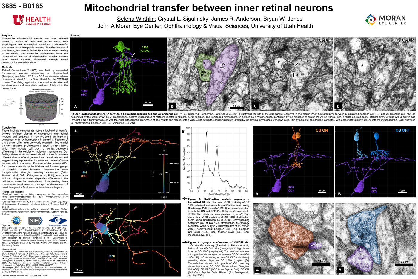
This abstract was presented today, April 26th at the 2023 Association for Research in Vision and Opthalmology (ARVO) meetings in New Orleans, Louisiana by Selena Wirthlin, Crystal Sigulinsky, James Anderson, and Bryan William Jones.

Full resolution version here.

Purpose
Intercellular mitochondrial transfer has been reported across a variety of cells and tissues under both physiological and pathological conditions. Such transfer has shown broad therapeutic potential. The effectiveness of this therapy, however, is limited by a lack of understanding of the cellular and molecular mechanisms. Here, the ultrastructural features of mitochondrial transfer between inner retinal neurons discovered through retinal connectomics analysis is shown.

Methods
Retinal Connectome 2 (RC2) was built by automated transmission electron microscopy at ultrastructural (2nm/pixel) resolution. RC2 is a 0.25mm diameter volume of retina obtained from a 5-month-old female C57BL/6J mouse. The Viking application was used to visualize and annotate inter- and intracellular features of interest in the connectome.

Results
Exploration of RC2 revealed material transfer between apposing neural processes within the OFF subliminal of the inner plexiform layer. The transferred material can be defined as a mitochondria, confirmed by the presence of crustae. At the transfer site, a short, electron-dense 140-nm diameter tube with a curved cap tightly associated with the inner mitochondrial membrane of one neuritis extends into a vacuole within the apposing neuritis formed by the plasma membranes of the two cells. Thin cytoskeletal components consistent with actin microfilaments extend into the mitochondrion. Morphology and synaptology of the acceptor cell confirm it is an Aii amacrine cell, while preliminary findings suggest the donor cell is a type of ON/OFF ganglion cell.

Conclusions
These findings demonstrate active mitochondrial transfer between different classes of endogenous inner retinal neurons and suggests it may represent an important component of tissue homeostasis in the retina. Features of this transfer differ from previously reported mitochondrial transfer between photoreceptors upon transplantation, which may indicate cell type- or context-dependent differences in the cellular or molecular mechanisms. Our findings demonstrate active mitochondrial transfer between different classes of endogenous inner retinal neurons and suggest it may represent an important component of tissue homeostasis in the retina. Features of this transfer differ from previous reports by the Wallace and Pearson groups of material transfer between photoreceptors upon transplantation through tunneling nanotubes (Ortin- Martinez et al., 2021; Kalargyrou et al., 2021), which may indicate cell type- or context-dependent differences in the cellular or molecular mechanisms. Understanding these mechanisms could serve as a catalyst for development of novel therapeutics for disease in the retina and beyond.

Müller Cell Connectomics In Health And Disease

This talk was presented today, April 25th at the 2023 Association for Research in Vision and Opthalmology (ARVO) meetings in New Orleans, Louisiana by Rebecca Pfeiffer as part of an ARVO Minisymposium Bryan William Jones organized.

Abstract: Muller cells are a critical component of retinal function and rapidly change metabolically and morphologically in retinal disease. Of Muller cell functions, many require close physical relationships between the Muller cell and the synapses of the neurons they support. Despite this required neuro-glial relationship, little is known about the direct contacts between Muller cells and synapses in healthy or diseased retinas. In order to address this, I use a connectomics/pathoconnectomics approach to reconstruct Muller cells and their neighboring synapses. The retinas evaluated are from a healthy rabbit, retinal connectome 1 (RC1), and from the P347L rabbit model of retinitis pigmentosa, retinal pathoconnectome 1 (RPC1). Preliminary data demonstrate an increase in endfoot entanglement in RPC1 when compared with RC1, and direct synaptic contact analysis of both connectomes is ongoing.

Species-Specific Connectivity In The Aii Connectome

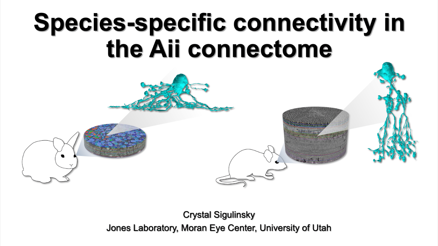
This talk was presented today, April 25th at the 2023 Association for Research in Vision and Opthalmology (ARVO) meetings in New Orleans, Louisiana by Crystal Sigulinsky as part of an ARVO Minisymposium organized by Bryan William Jones.

Abstract: Biomedical research relies heavily on animal models to study human disease and develop therapeutics. Understanding the architectural diversity in neural networks between humans and these model species is essential for choosing a relevant study model and interpreting conflicting results. Using comparative connectomics, we sought to map and compare the local neural network architecture of rabbit and mouse retinal Aii amacrine cells. This specialized narrow-field, multistratified, glycinergic interneuron has critical feedforward and feedback roles in both the photopic and scotopic retinal networks spanning the ON and OFF pathways, making it an ideal candidate for investigating species-specific differences in retinal networks. High-resolution, serial-section transmission electron microscopy (TEM) volumes of rabbit (RC1: female, 13- month, Dutch Belted) and mouse (RC2: female, 5-month, C57BL/6J) retinal tissue provided spatially-registered synaptic maps of Aii connectivity at directly comparable resolution and completeness. These reveal that despite species-specific morphologies, gross synaptology and compartmentalization appear conserved. Yet, rabbit and mouse Aii cells diverge in the weighting of their partnerships, most notably in their coupling profiles. Opposing biases in gap junction partnerships and their respective sizing rules indicate a greater relative output by mouse Aii cells to ON pathways than in rabbit. However, a unique topological conformation for a subset of conventional presynapses formed by Aii cell lobular dendrites with species-specific features and prevalence may influence signal output to specific partner classes within the OFF pathway and either nullify or exacerbate this difference in ON/OFF output. Additionally, rabbit Aii cells in RC1 showed greater Aii-Aii coupling than in mouse, which may suggest greater signal-to-noise compensation. Lastly, preliminary data suggest mouse Aii cells receive greater excitatory, but not inhibitory input/feedback from the OFF pathway than in rabbit. Together these data indicate that precise neural circuit architectures diverge between species and require detailed, comprehensive mapping to begin to dissect potential influence on signal flow.

Structural Motifs Of Excitatory Synapses In The Mammalian Retina

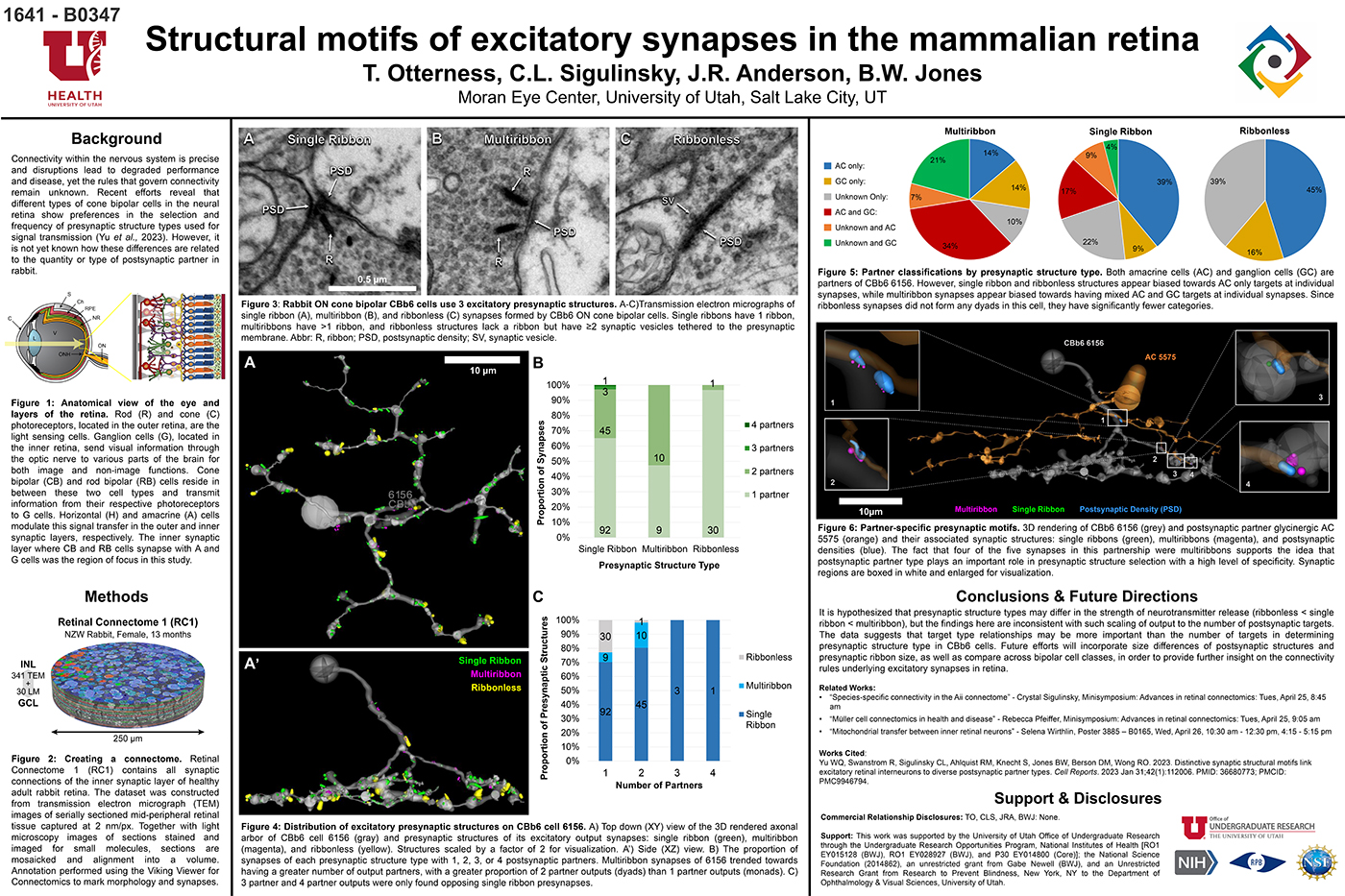
This abstract was presented today, April 24th at the 2023 Association for Research in Vision and Opthalmology (ARVO) meetings in New Orleans, Louisiana by Taylor Otterness, Crystal Sigulinsky, James Anderson, and Bryan William Jones.

Full resolution version here.

Purpose
Connectivity within the nervous system is precise and disruptions lead to degraded performance and disease, yet the rules that govern connectivity remain unknown. Recent efforts reveal that different types of cone bipolar cells in the neural retina show preferences in the selection and frequency of presynaptic structure types used for signal transmission. However, it is not yet known how these differences are related to the quantity or type of postsynaptic partner. We used Retinal Connectome 1 (RC1) to analyze the synaptic output of rabbit CBb6 cells, a type of ON cone bipolar cell that forms excitatory synapses via diverse presynaptic structure types, to identify patterns in how these cells interact with their postsynaptic partners.

Methods
RC1 is a 0.25 mm diameter volume sampled from mid-peripheral retina of a 13 month old female Dutch-Belted rabbit, serially sectioned at 70 nm, and imaged at ultrastructural resolution (2nm/px) using transmission electron microscopy. Postsynaptic partners of CBb6 cell 6156’s presynaptic structures were annotated using the Viking Viewer for Connectomics. Statistical analyses were conducted in Microsoft Excel and investigated further with 3D rendering and graph visualization of connectivity.

Results
The factors tracked for comparison included presynaptic structure type, target number, and postsynaptic partner type. Multiribbon synapses of CBb6 cell 6156 trended towards having a greater number of output partners, with a greater proportion of dyads than monads. Despite this, triads and quadrads were only found opposing single ribbon synapses. As the different presynaptic structure types may differ in the strength of neurotransmitter release (ribbonless < single ribbon < multiribbon), these findings are inconsistent with scaling of output to the number of postsynaptic targets. Both amacrine cells (AC) and ganglion cells (GC) are postsynaptic partners of 6156. However, single ribbon and ribbonless structures appear biased towards AC only targets, while multiribbon synapses appear biased toward mixed AC and GC targets.

Conclusions
Target type relationships appear more important than the number of targets in determining presynaptic structure type in CBb6. Future efforts will examine size differences of postsynaptic structures and presynaptic ribbon size, and even compare across bipolar cell classes, in order to provide further insight on the connectivity rules underlying excitatory synapses.

Distinctive Synaptic Structural Motifs Link Excitatory Retinal Interneurons To Diverse Postsynaptic Partner Types

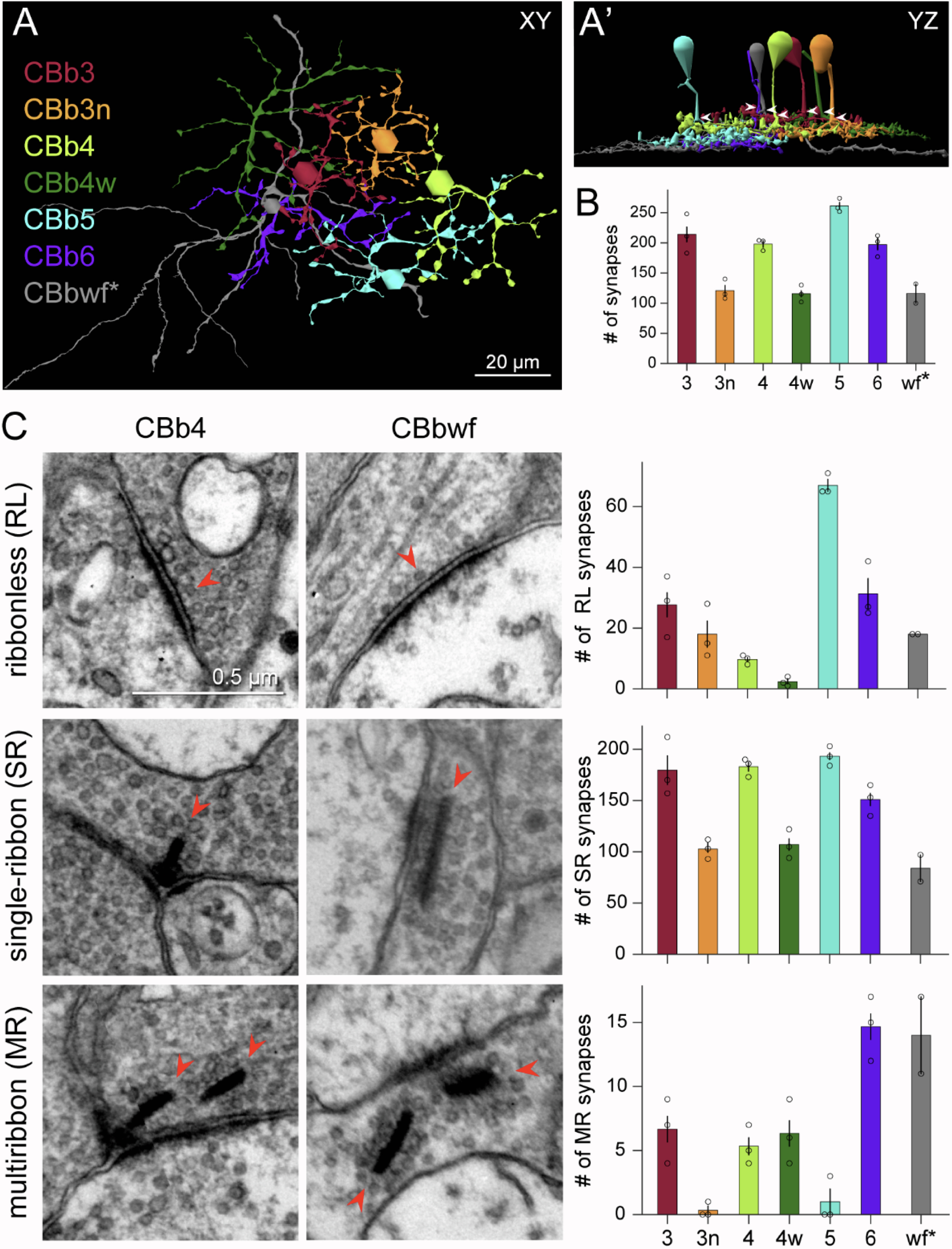
We have a new manuscript from the lab in Cell Reports, Distinctive Synaptic Structural Motifs Link Excitatory Retinal Interneurons To Diverse Postsynaptic Partner Types. This manuscript is in collaboration with the first author, Wan-Qing Yu @wanqing_yu, then co-authors Rachael Swanstrom, Crystal L. Sigulinsky @CSigulinsky, Richard M. Ahlquist, along with Sharm Knecht, myself Bryan W. Jones @BWJonesDavid M. Berson, and Rachel O. Wong. The PDF is here.

Abstract:
Neurons make converging and diverging synaptic connections with distinct partner types. Whether synapses involving separate partners demonstrate similar or distinct structural motifs is not yet well understood. We thus used serial electron microscopy in mouse retina to map output synapses of cone bipolar cells (CBCs) and compare their structural arrangements across bipolar types and postsynaptic partners. Three presynaptic configurations emerge—single-ribbon, ribbonless, and multiribbon synapses. Each CBC type exploits these arrangements in a unique combination, a feature also found among rabbit ON CBCs. Though most synapses are dyads, monads and triads are also seen. Altogether, mouse CBCs exhibit at least six motifs, and each CBC type uses these in a stereotypic pattern. Moreover, synapses between CBCs and particular partner types appear biased toward certain motifs. Our observations reveal synaptic strategies that diversify the output within and across CBC types, potentially shaping the distinct functions of retinal microcircuits.

Current Perspective on Retinal Remodeling: Implications for Therapeutics

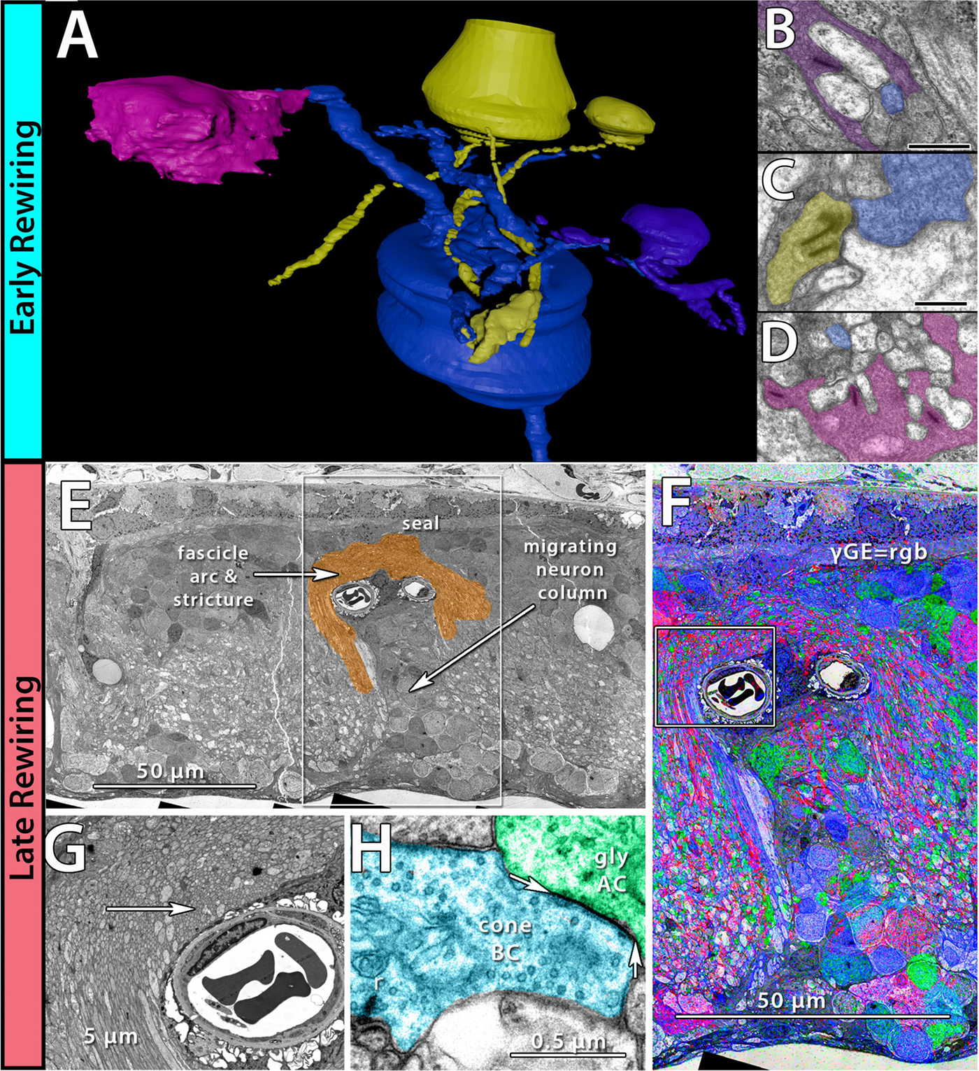
We have a new paper out of the lab, a perspectives paper on Retinal Remodeling: Implications for Therapeutics. (pdf here).

Authors are Rebecca L. Pfeiffer @BeccaPfeiffer19, and Bryan W. Jones @BWJones.

Abstract: The retinal degenerative diseases retinitis pigmentosa and age-related macular degeneration are a leading cause of irreversible vision loss. Both present with progressive photoreceptor degeneration that is further complicated by processes of retinal remodeling. In this perspective, we discuss the current state of the field of retinal remodeling and its implications for vision-restoring therapeutics currently in development. Here, we discuss the challenges and pitfalls retinal remodeling poses for each therapeutic strategy under the premise that understanding the features of retinal remodeling in totality will provide a basic framework with which therapeutics can interface. Additionally, we discuss the potential for approaching therapeutics using a combined strategy of using diffusible molecules in tandem with other vision-restoring therapeutics. We end by discussing the potential of the retina and retinal remodeling as a model system for more broadly understanding the progression of neurodegeneration across the central nervous system.

Revival Of Light Signalling In The Postmortem Mouse And Human Retina

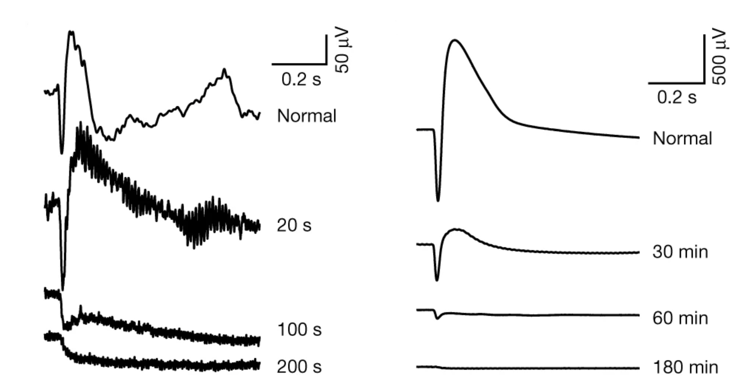
We have a new collaborative manuscript out in Nature, Revival of light signalling in the postmortem mouse and human retina. Full paper (here).

Authors: Fatima Abbas @neurofim, Silke Becker, Bryan W. Jones @BWJones, Ludovic S. Mure, Satchidananda Panda @SatchinPanda, Anne Hanneken & Frans Vinberg @fvinberg.

Abstract:
Death is defined as the irreversible cessation of circulatory, respiratory or brain activity. Many peripheral human organs can be transplanted from deceased donors using protocols to optimize viability. However, tissues from the central nervous system rapidly lose viability after circulation ceases, impeding their potential for transplantation. However, the time course and mechanisms causing neuronal death and the potential for revival remain poorly defined. Here, using the retina as a model of the central nervous system, we systemically examine the kinetics of death and neuronal revival. We demonstrate the swift decline of neuronal signalling and identify conditions for reviving synchronous in vivo-like trans-synaptic transmission in postmortem mouse and human retina. We measure light-evoked responses in human macular photoreceptors in eyes removed up to 5 h after death and identify modifiable factors that drive reversible and irreversible loss of light signalling after death. Finally, we quantify the rate-limiting deactivation reaction of phototransduction, a model G protein signalling cascade, in peripheral and macular human and macaque retina. Our approach will have broad applications and impact by enabling transformative studies in the human central nervous system, raising questions about the irreversibility of neuronal cell death, and providing new avenues for visual rehabilitation.

Model-Based Comparison of Current Flow in Rod Bipolar Cells of Healthy and Early-Stage Degenerated Retina

We have a new manuscript out in Experimental Eye Research, Model-Based Comparison of Current Flow in Rod Bipolar Cells of Healthy and Early-Stage Degenerated Retina. (pdf here)

Authors: Pragya Kosta, Ege Iseri, Kyle Loizos, Javad Paknahad, Rebecca L. Pfeiffer @BeccaPfeiffer19, Crystal L. Sigulinsky @CLSigulinsky, James R. Anderson, Bryan W. Jones @BWJones, and Gianluca Lazzi.

Abstract: Retinal degenerative diseases, such as retinitis pigmentosa, are generally thought to initiate with the loss of photoreceptors, though recent work suggests that plasticity and remodeling occurs prior to photoreceptor cell loss. This degeneration subsequently leads to death of other retinal neurons, creating functional alterations and extensive remodeling of retinal networks. Retinal prosthetic devices stimulate the surviving retinal cells by applying external current using implanted electrodes. Although these devices restore partial vision, the quality of restored vision is limited. Further knowledge about the precise changes in degenerated retina as the disease progresses is essential to understand how current flows in retinas undergoing degenerative disease and to improve the performance of retinal prostheses. We developed computational models that describe current flow from rod photoreceptors to rod bipolar cells


Model-based Comparison of Current Flow in Rod Bipolar Cells of Healthy and Early-Stage Degenerated Retina

Contrast Sensitivity Isocontours Of The Central Visual Field

We have a new manuscript out in Nature Scientific Reports, Contrast Sensitivity Isocontours Of The Central Visual Field.

Authors: Agnes Y. J. Choi, Lisa Nivison-Smith, Jack Phu, Barbara Zangerl, Sieu K. Khuu, Bryan W. Jones, Rebecca L. Pfeiffer, Robert E. Marc & Michael Kalloniatis.

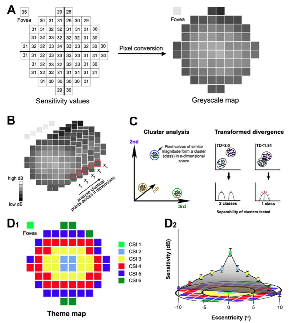
Abstract: Standard automated perimetry (SAP), the most common form of perimetry used in clinical practice, is associated with high test variability, impacting clinical decision making and efficiency. Contrast sensitivity isocontours (CSIs) may reduce test variability in SAP by identifying regions of the visual field with statistically similar patterns of change that can be analysed collectively and allow a point (disease)-to-CSI (normal) comparison in disease assessment as opposed to a point (disease)-to-point (normal) comparison. CSIs in the central visual field however have limited applicability as they have only been described using visual field test patterns with low, 6° spatial sampling. In this study, CSIs were determined within the central 20° visual field using the 10-2 test grid paradigm of the Humphrey Field Analyzer which has a high 2° sampling frequency. The number of CSIs detected in the central 20° visual field was greater than previously reported with low spatial sampling and stimulus size dependent: 6 CSIs for GI, 4 CSIs for GII and GIII, and 3 CSIs for GIV and GV. CSI number and distribution were preserved with age. Use of CSIs to assess visual function in age-related macular degeneration (AMD) found CSI guided analysis detected a significantly greater deviation in sensitivity of AMD eyes from normal compared to a standard clinical pointwise comparison (−1.40 ± 0.15 dB vs −0.96 ± 0.15 dB; p < 0.05). This work suggests detection of CSIs within the central 20° is dependent on sampling strategy and stimulus size and normative distribution limits of CSIs can indicate significant functional deficits in diseases affecting the central visual field such as AMD.

Coupling architecture of the retinal Aii/ON cone bipolar cell network and alteration in degeneration

This poster was presented today, July 28th at the 2019 International Gap Junction Conference in Victoria, Canada by Crystal L. Sigulinsky, Rebecca L. PfeifferJames R. Anderson, Christopher Rapp, Jeebika Dahal, Jessica C Garcia, Jia-Hui Yang, Daniel P. Emrich, Hope Morrison, Kevin D. Rapp, Carl B. Watt, Mineo Kondo, Hiroko Terasaki, Robert E. Marc and Bryan W. Jones.

Almost full resolution version here.

Authors:

Crystal L Sigulinsky1, Rebecca L Pfeiffer1, James R. Anderson1, Christopher N. Rapp1, Jeebika Dahal1, Jessica C Garcia1, Jia-Hui Yang1, Daniel P. Emrich1, Hope Morrison1, Kevin D. Rapp1, Carl B. Watt1, Mineo Kondo2, Hiroko Terasaki3, Robert E. Marc1, Bryan W. Jones1
1Moran Eye Center/ Ophthalmology, University of Utah, Salt Lake City, Utah, United States; 2Mie University, Tsu, Japan; 3Nagoya University, Nagoya-shi, Japan;

ABSTRACT:

Background and aim:
Gap junctions are prevalent throughout the neural retina, with expression by every major neuronal class and at every level of signal processing. Yet, the functional roles and expressing cells/participating networks for many remain unknown. Spontaneous network spontaneous hyperactivity observed during retinal degeneration contributes to visual impairment and requires gap junctional coupling in the Aii amacrine cell/ON cone bipolar cell (CBC) network.  However, it remains unclear whether this hyperactivity reflects changes in the underlying circuitry or dysfunction of the normative circuitry. Here, we used connectomics-based mapping of retinal circuitry to 1) define the coupling architecture of the Aii/ON CBC network in healthy adult rabbit retina using connectome RC1 and 2) evaluate changes in coupling motifs in RPC1, a pathoconnectome from a rabbit retinal degeneration model.

Methods:

RC1 and RPC1 are connectomes built by automated transmission electron microscopy at ultrastructural (2 nm/pixel) resolution. RC1 is a 0.25 mm diameter volume of retina from a 13-month old, light adapted female Dutch Belted rabbit. RPC1 is a 0.07 mm diameter volume of degenerate retina from a transgenic P347L model of autosomal dominant retinitis pigmentosa (10-months old, male, New Zealand White background) presenting with ~50% rod loss. ON CBCs, Aii amacrine cells, and their coupling partners were annotated using the Viking application. Coupling motifs and features were explored with 3D rendering and network graph visualization. Gap junctions were validated by 0.25 nm resolution recapture with goniometric tilt when necessary.

Results:

Complete reconstruction of 37 ON CBCs in RC1 yielded 1339 gap junctions and revealed pervasive in- and cross-class coupling motifs among ON CBCs that produce complex network topologies within the coupled Aii network. Robust rulesets underlie class-specific coupling profiles with specificity defined beyond geometric opportunity. These coupling profiles enabled classification of all 145 ON CBCs contained within RC1 into 7 distinct classes. In RPC1, two ON CBC classes appear to retain their class-specific coupling profiles, accepting and rejecting specific combinations of Aii and ON CBC class partnerships. However, aberrant partnerships exist, including both loss of motifs and acquisition of novel ones.

Conclusions:

Gap junctions formed by ON CBCs are prominent network components, with specificity rivaling that of chemical synapses. These gap junctions not only subserve canonical signal transfer for night vision, but also extensive coupling within and across the parallel processing streams. Clearly aberrant morphological and synaptic changes exist in RPC1, including changes in the coupling specificity of both Aii and ON CBCs. Thus, circuit topology is altered prior to complete loss of rods, with substantial implications for therapeutic interventions for blinding diseases that depend upon the surviving retinal network.