Tag Archives: Rebecca Pfeiffer

Single-Cell Profiling of Trabecular Meshwork Identifies Mitochondrial Dysfunction In a Glaucoma Model That Is Protected By Vitamin B3 Treatment


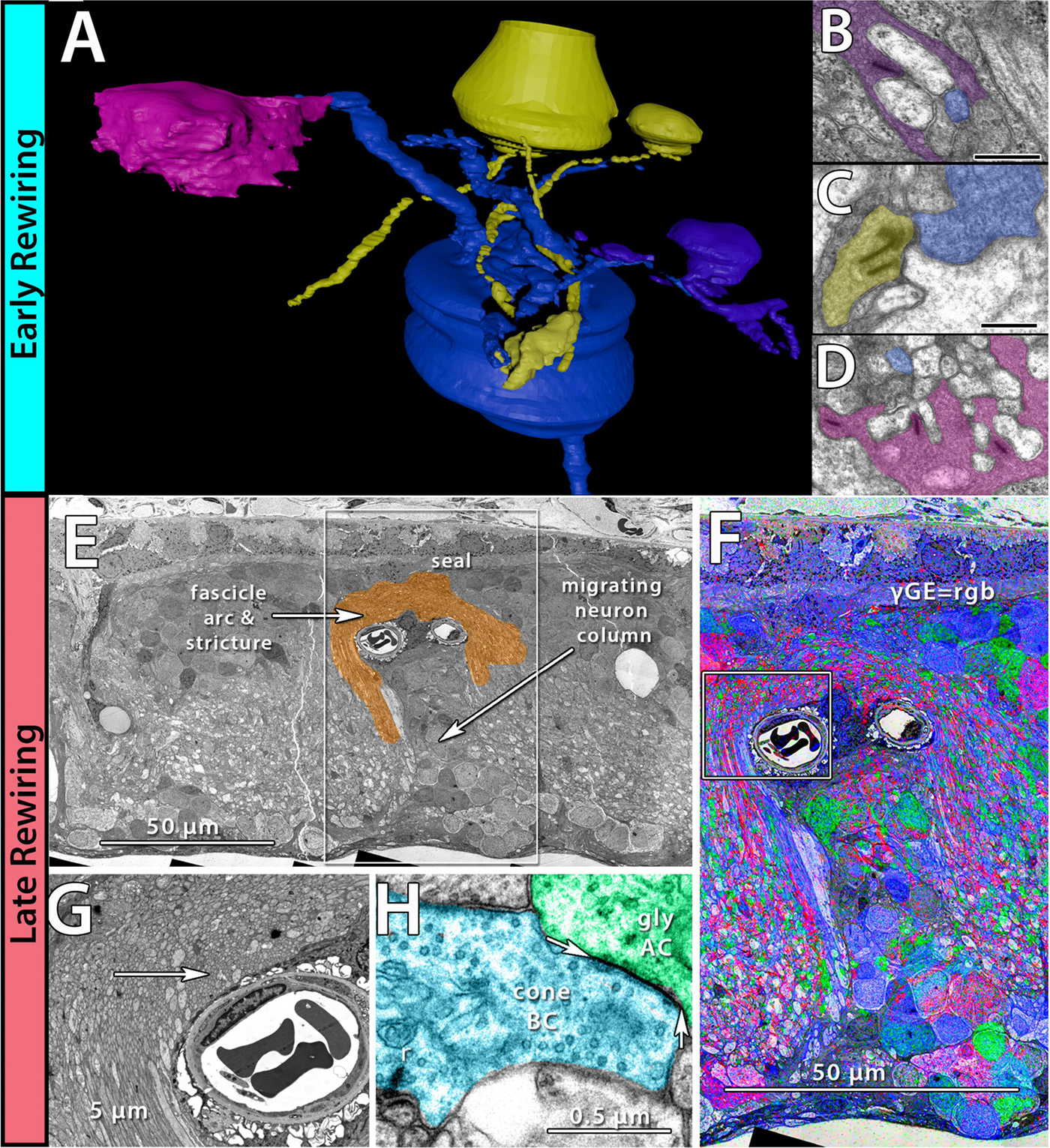
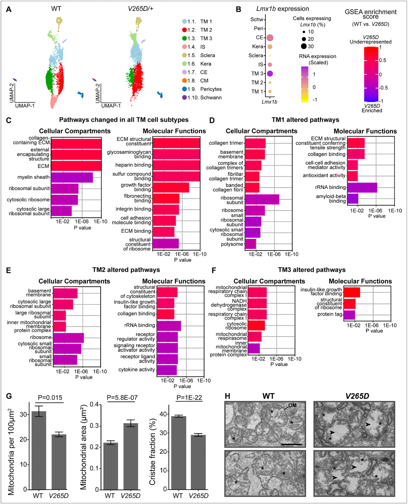
We have a new collaborative paper out in eLife: Single-Cell Profiling of Trabecular Meshwork Identifies Mitochondrial Dysfunction In a Glaucoma Model That Is Protected By Vitamin B3 Treatment.

Authors: Nicholas Tolman, Taibo Li, Revathi Balasubramanian, Guorong Li, Rebecca L. Pfeiffer@beccapfeiffer.bsky.social, Violet Bupp-Chickering, Ruth A Kelly, Marina Simón, John Peregrin, Christa Montgomery, Bryan Jones @bwjones.bsky.social, W Daniel Stamer, Jiang Qian, Simon WM John

Abstract: Since the trabecular meshwork (TM) is central to intraocular pressure (IOP) regulation and glaucoma, a deeper understanding of its genomic landscape is needed. We present a multi-modal, single-­ cell resolution analysis of mouse limbal cells (includes TM). In total, we sequenced 9,394 wild-­ type TM cell transcriptomes. We discovered three TM cell subtypes with characteristic signature genes validated by immunofluorescence on tissue sections and whole-­ mounts. The subtypes are robust, being detected in datasets for two diverse mouse strains and in independent data from two institutions. Results show compartmentalized enrichment of critical pathways in specific TM cell subtypes. Distinctive signatures include increased expression of genes responsible for (1) extracellular matrix structure and metabolism (TM1 subtype), (2) secreted ligand signaling to support Schlemm’s canal cells (TM2), and (3) contractile and mitochondrial/metabolic activity (TM3). ATAC-­ sequencing data identified active transcription factors in TM cells, including LMX1B. Mutations in LMX1B cause high IOP and glaucoma. LMX1B is emerging as a key transcription factor for normal mitochondrial function, and its expression is much higher in TM3 cells than other limbal cells. To understand the role of LMX1B in TM function and glaucoma, we single-­ cell sequenced limbal cells from Lmx1bV265D/+mutant mice (2491 TM cells). In Lmx1bV265D/+ mice, TM3 cells were uniquely affected by pronounced mitochondrial pathway changes. Mitochondria in TM cells of Lmx1bV265D/+ mice are swollen with a reduced cristae area, further supporting a role for mitochondrial dysfunction in the initiation of IOP elevation in these mice. Importantly, treatment with vitamin B3 (nicotinamide), which enhances mitochondrial function and metabolic resilience in other contexts, significantly protected Lmx1b mutant mice from IOP elevation.

Current Perspective on Retinal Remodeling: Implications for Therapeutics

We have a new paper out of the lab, a perspectives paper on Retinal Remodeling: Implications for Therapeutics. (pdf here).

Authors are Rebecca L. Pfeiffer @BeccaPfeiffer19, and Bryan W. Jones @BWJones.

Abstract: The retinal degenerative diseases retinitis pigmentosa and age-related macular degeneration are a leading cause of irreversible vision loss. Both present with progressive photoreceptor degeneration that is further complicated by processes of retinal remodeling. In this perspective, we discuss the current state of the field of retinal remodeling and its implications for vision-restoring therapeutics currently in development. Here, we discuss the challenges and pitfalls retinal remodeling poses for each therapeutic strategy under the premise that understanding the features of retinal remodeling in totality will provide a basic framework with which therapeutics can interface. Additionally, we discuss the potential for approaching therapeutics using a combined strategy of using diffusible molecules in tandem with other vision-restoring therapeutics. We end by discussing the potential of the retina and retinal remodeling as a model system for more broadly understanding the progression of neurodegeneration across the central nervous system.

Proteomic changes in the lens of a congenital cataract mouse model lead to reduced levels of glutathione and taurine

This abstract was presented today, May 4th at the 2022  Association for Research in Vision and Opthalmology (ARVO) meetings in Denver, Colorado by Sheldon Rowan @SheldonRowan, Eloy Bejarano, Elizabeth Whitcomb, Rebecca Pfeiffer @BeccaPfeiffer19, Kristie Rose, Kevin Schey, Bryan Jones @BWJones, Allen Taylor.

Purpose: Congenital cataracts develop through multiple mechanisms, but often lead to common endpoints, including protein aggregation, impaired fiber cell differentiation, and absence of fiber cell denucleation. It is now apparent that other metabolic abnormalities associate with cataractogenesis, including reductions in levels of amino acids, glutathione, and taurine. Here, we analyze the proteome and metabolome of mice expressing a mutant ubiquitin protein (K6W-Ub) to determine the molecular mechanisms underlying formation of its congenital cataract.

Methods: C57BL/6J wild-type or cataractous K6W-Ub transgenic mouse lenses were dissected at E15.5, P1, or P30 and proteins were analyzed via MS-based tandem-mass-tag (TMT) quantitative proteomics. Small molecules were spatially quantified using computational molecular phenotyping (CMP), a tool that enables acquisition of free amino acid fingerprints for every cell in the lens. Validation of proteomics findings was also performed using Western blot analysis and immunohistochemistry.

Results: Proteomic analyses revealed pathways that were altered during lens differentiation, by expression of K6W-Ub, or both. Prominent pathways included glutathione metabolism; glycolysis/gluconeogenesis; and glycine, serine, and threonine metabolism. Within the glutathione metabolism pathway, GSTP1 and GGCT were most strongly downregulated by K6W-Ub. Other consistently downregulated proteins were PGAM2, GAMT, and HMOX1. Proteins that were upregulated by K6W-Ub expression belonged to pathways related to lysosome, autophagy, Alzheimer’s disease, and glycolysis/gluconeogenesis. Analysis of the metabolome via CMP revealed statistically significant decreases in taurine and glutathione and smaller decreases in glutamate, glutamine, aspartate, and valine in all ages of K6W-Ub lenses. Lens metabolites were spatially altered in the cataractous K6W-Ub lens.

Conclusions: K6W-Ub expressing lenses replicate many congenital cataract phenotypes and are useful disease models. The large reductions in levels of taurine and glutathione may be general signatures of cataract development, as human cataracts also have reduced glutathione and taurine. Key roles for amino acid metabolism and glycolysis/gluconeogenesis in cataractogenesis are emerging. Together our data point toward potential common metabolic/proteomic signatures of cataracts.

Becca Pfeiffer At TEM2

This is Becca Pfeiffer setting up a test capture on our new JEOL electron microscope. We’ve customized this scope, like our previous scope, and it is taking us a little while to track down some variables with a piece of equipment this complex and hammer them down. My thanks to Becca, Jamie and Kevin for working through this together.

From Jonesblog.

Pathoconnectome Analysis of Müller Cells in Early Retinal Remodeling

Rebecca Pfeiffer, a post-doc in the laboratory presented her work on “Pathoconnectome Analysis of Müller Cells in Early Retinal Remodeling” as a platform presentation at the RD2018 meeting in Killarney, Ireland.

Authors: Rebecca Pfeiffer, James R. Anderson, Daniel P. Emrich, Jeebika Dahal, Crystal L Sigulinsky, Hope AB Morrison, Jia-Hui Yang, Carl B. Watt, Kevin D. Rapp, Mineo Kondo, Hiroko Terasaki, Jessica C Garcia, Robert E. Marc, and Bryan W. Jones.

Purpose: Glia play important roles in neural system function. These roles include, but are not limited to: amino acid recycling, ion homeostasis, glucose transport, and removal of waste. During retinal degeneration, Muller cells, the primary macroglia of the retina, are one of the first cells to show metabolic and morphological alterations in response to retinal stress. The metabolic alterations observed in Muller cells appear to manifest in regions of photoreceptor degeneration; however, the precise mechanisms that govern these alterations in response to neuronal stress, synapse maintenance, or glia-glia interactions is currently unknown.  This project aims to reconstruct Muller cells from a pathoconnectome of early retinal remodeling at 2nm/pixel with ultrastructural metabolic data to determine the relationship of structural and metabolic phenotypes between neighboring neurons and glia.

Methods:  Retinal pathoconnectome 1 (RPC1) is the first connectome to be assembled from pathologic neural tissue (a pathoconnectome). The tissue selected for RPC1 was collected post mortem from a 10 month transgenic P347L rabbit model of autosomal dominant retinitis pigmentosa, fixed in 1% formaldehyde, 2.5% glutaraldehyde, 3% sucrose, and 1mM MgSO4 in cacodylate buffer (pH 7.4). The tissue was subsequently osmicated, dehydrated, resin embedded, and sectioned at 70nm. Sections were placed on formvar grids, stained, and imaged at 2nm/pixel on a JEOL JEM-1400 TEM using SerialEM software. 1 section was reserved from every 30 sections for CMP, where it was placed on a slide and probed for small molecules: glutamate, glutamine, glycine, GABA, taurine, glutathione; or TEM compatible proteins GFAP and GS. The pathoconnectome volume was evaluated and annotated using the Viking software suite.

Results: RPC1 demonstrates hallmarks of early retinal degeneration and remodeling, including the glial phenotypes of hypertrophy and metabolic variation between neighboring Muller cells. Early evaluation of these glia demonstrates variations in osmication in Muller cells as well as apparent encroachment of glial end-feet on one another.  We are currently in the process of reconstructing multiple Muller cells within RPC1 and their neighboring neurons.  Once complete, we will assess the relationship between Muller cell phenotype and the phenotypes of contacted neuronal and glial neighbors.

Conclusions: How neural-glial relationships are affected by retinal remodeling may help us understand why remodeling and neurodegeneration follow photoreceptor degeneration. In addition, determining these relationships during remodeling will be crucial to developing therapeutics with long-term success. RPC1 provides a framework to analyze these relationships in early retinal remodeling through ultrastructural reconstructions of all neurons and glia in an intact retina. These reconstructions, informed by quantitative metabolite labeling, will allow us to evaluate these neural-glial interactions more comprehensively than other techniques have previously allowed.

Off to RD2018 and ISER 2018

The Marclab for Connectomics is off to RD2018 and ISER 2018 in Killarney, Ireland and Belfast, Northern Ireland.  I’ll be organizing sessions on retinal degeneration, and I’m tremendously proud of the work Dr. Crystal Sigulinsky will be presenting from her work on gap junctional connectivity in retinal degenerations and the work Dr. Rebecca Pfeiffer (@BeccaPfeiffer19) will be presenting on her work on the retinal pathoconnectome in two talks on bipolar cells and Müller cells.

A Pathoconnectome of Early Retinal Remodeling

This abstract was presented today, Monday, April 30th at the 2018 Association for Research in Vision and Opthalmology (ARVO) meetings in Honolulu, Hawaii by Rebecca Pfeiffer, Robert E. Marc, James R. Anderson, Daniel P. Emrich, Carl B. Watt, Jia-Hui Yang, Kevin D. Rapp, Jeebika Dahal, Mineo Kondo, Hiroko Terasaki, and Bryan W. Jones.

Purpose:
Retinal remodeling is a consequence of retinal degenerative disease, during which neurons sprout new neurites whose synaptic structures and partners are not yet defined. Simultaneously during remodeling, Müller cells (MCs) undergo structural and metabolic changes, whose impact on surrounding neurons is an active area of research. Retinal connectomes have elucidated and validated fundamental networks. These data provide further classification of neuronal types and subtypes and a precise framework for modeling of retinal function, based on ground truth networks. The creation of the first pathoconnectome (RPC1), a connectome from pathological retinal tissue, provides the opportunity to determine connectivites between neurons, while simultaneously evaluating glial remodeling. Computational Molecular Phenotyping (CMP) embedded within the ultrastructure provides metabolic factors of pathologies.

Methods:
RPC1 was collected post-mortem from a 10mo TgP347L rabbit model of adRP, fixed in 1% FA, 2.5% GA, 3% sucrose, and 1mM MgSO4 in cacodylate buffer (pH 7.4). The tissue was osmicated, dehydrated, resin embedded, and sectioned at 70nm. Sections were placed on formvar grids, stained, and imaged on a JEOL JEM-1400 TEM using SerialEM. 1 section was reserved from every 30 section for CMP, where it was probed for small molecules: glutamate, glutamine, glycine, GABA, taurine, glutathione; or proteins GFAP and GS. RPC1 was evaluated using the Viking software suite.

Results:
RPC1 was chosen based on early features of retinal degeneration/remodeling: degeneration of rod OS, MC hypertrophy, and neuronal sprouting. RPC1 consists of 948 serial sections spanning the ONL to the vitreous, with a diameter of 90µm. We find dendrites extending from rod bipolar cells to cone pedicles, originally described in light microscopy, and active synaptic contacts. We also see alterations of synaptic structure in the IPL, and MC morphological changes affecting surface to volume and neuron/glial relationships. Network motifs are being actively investigated.

Conclusions:
We observe many features of remodeling previously described using light microscopy, and confirm active synaptic contact. We also find synaptic structural features, not previously described. In addition, early evaluation of MC morphology demonstrates marked changes in MC shape and associations with nearby neurons and glia, which, combined with CMP, will be instrumental in understanding how MCs affect retinal remodeling.

Pattern Recognition Analysis Reveals Unique Contrast Sensitivity Isocontours Using Static Perimetry Thresholds Across The Visual Field

We have a new publication in IOVS, Pattern Recognition Analysis Reveals Unique Contrast Sensitivity Isocontours Using Static Perimetry Thresholds Across The Visual Field (Direct link here).  Authors are:  Jack Phu, Sieu Khuu, Lisa Nivison-Smith, Barbara Zangerl, Agnes Yiu, Jeung Choi, Bryan W. JonesRebecca Pfeiffer, Robert Marc, and Michael Kalloniatis.

Purpose
To determine the locus of test locations that exhibit statistically similar age-related decline in sensitivity to light increments and age-corrected contrast sensitivity isocontours (CSIs) across the central visual field (VF). We compared these CSIs with test point clusters used by the Glaucoma Hemifield Test (GHT).

Methods
Sixty healthy observers underwent testing on the Humphrey Field Analyzer 30-2 test grid using Goldmann (G) stimulus sizes I-V. Age-correction factors for GI-V were determined using linear regression analysis. Pattern recognition analysis was used to cluster test locations across the VF exhibiting equal age-related sensitivity decline (age-related CSIs), and points of equal age-corrected sensitivity (age-corrected CSIs) for GI-V.

Results
There was a small but significant test size–dependent sensitivity decline with age, with smaller stimuli declining more rapidly. Age-related decline in sensitivity was more rapid in the periphery. A greater number of unique age-related CSIs was revealed when using smaller stimuli, particularly in the mid-periphery. Cluster analysis of age-corrected sensitivity thresholds revealed unique CSIs for GI-V, with smaller stimuli having a greater number of unique clusters. Zones examined by the GHT consisted of test locations that did not necessarily belong to the same CSI, particularly in the periphery.

Conclusions
Cluster analysis reveals statistically significant groups of test locations within the 30-2 test grid exhibiting the same age-related decline. CSIs facilitate pooling of sensitivities to reduce the variability of individual test locations. These CSIs could guide future structure-function and alternate hemifield asymmetry analyses by comparing matched areas of similar sensitivity signatures.