Single-Cell Profiling of Trabecular Meshwork Identifies Mitochondrial Dysfunction In a Glaucoma Model That Is Protected By Vitamin B3 Treatment


We have a new collaborative paper out in eLife: Single-Cell Profiling of Trabecular Meshwork Identifies Mitochondrial Dysfunction In a Glaucoma Model That Is Protected By Vitamin B3 Treatment.

Authors: Nicholas Tolman, Taibo Li, Revathi Balasubramanian, Guorong Li, Rebecca L. Pfeiffer@beccapfeiffer.bsky.social, Violet Bupp-Chickering, Ruth A Kelly, Marina Simón, John Peregrin, Christa Montgomery, Bryan Jones @bwjones.bsky.social, W Daniel Stamer, Jiang Qian, Simon WM John

Abstract: Since the trabecular meshwork (TM) is central to intraocular pressure (IOP) regulation and glaucoma, a deeper understanding of its genomic landscape is needed. We present a multi-modal, single-­ cell resolution analysis of mouse limbal cells (includes TM). In total, we sequenced 9,394 wild-­ type TM cell transcriptomes. We discovered three TM cell subtypes with characteristic signature genes validated by immunofluorescence on tissue sections and whole-­ mounts. The subtypes are robust, being detected in datasets for two diverse mouse strains and in independent data from two institutions. Results show compartmentalized enrichment of critical pathways in specific TM cell subtypes. Distinctive signatures include increased expression of genes responsible for (1) extracellular matrix structure and metabolism (TM1 subtype), (2) secreted ligand signaling to support Schlemm’s canal cells (TM2), and (3) contractile and mitochondrial/metabolic activity (TM3). ATAC-­ sequencing data identified active transcription factors in TM cells, including LMX1B. Mutations in LMX1B cause high IOP and glaucoma. LMX1B is emerging as a key transcription factor for normal mitochondrial function, and its expression is much higher in TM3 cells than other limbal cells. To understand the role of LMX1B in TM function and glaucoma, we single-­ cell sequenced limbal cells from Lmx1bV265D/+mutant mice (2491 TM cells). In Lmx1bV265D/+ mice, TM3 cells were uniquely affected by pronounced mitochondrial pathway changes. Mitochondria in TM cells of Lmx1bV265D/+ mice are swollen with a reduced cristae area, further supporting a role for mitochondrial dysfunction in the initiation of IOP elevation in these mice. Importantly, treatment with vitamin B3 (nicotinamide), which enhances mitochondrial function and metabolic resilience in other contexts, significantly protected Lmx1b mutant mice from IOP elevation.

Uncovering the electrical synapse proteome in retinal neurons via in vivo proximity labeling

We have a new paper out in eLife: Uncovering the electrical synapse proteome in retinal neurons via in vivo proximity labeling.

Authors: Stephan Tetenborg, Eyad Shihabeddin, Elizebeth Olive Akansha Manoj Kumar, Crystal Sigulinsky @csigulinsky.bsky.social, Karin Dedek, Ya-Ping Lin, Fabio Echeverry, Hannah Hoff, Alberto Pereda, Bryan William Jones @bwjones.bsky.social, Christophe Ribelayga, Klaus Ebnet, Ken Matsuura, John O’Brien

Abstract: Electrical synapses containing Connexin 36 (Cx36) represent the main means for direct electrical communication among neurons in the mammalian nervous system. However, little is known about the protein complexes that constitute these synapses. In the present study, we applied different BioID strategies to screen the interactomes of Connexin 36 and its zebrafish orthologue Cx35b in retinal neurons. For in vivo proximity labeling in mice, we took advantage of the Cx36-EGFP strain and expressed a GFP-nanobody-TurboID fusion construct selectively in AII amacrine cells. For in vivo BioID in zebrafish, we generated a transgenic line expressing a Cx35b-TurboID fusion under control of the Cx35b promoter. Both strategies allowed us to capture a plethora of molecules that were associated with electrical synapses and showed a high degree of evolutionary conservation in the proteomes of both species. Besides known interactors of Cx36 such as ZO-1 and ZO-2 we have identified more than 50 new proteins, such as scaffold proteins, adhesion molecules and regulators of the cytoskeleton. Moreover, we determined the subcellular localization of these proteins in mouse retina and tested potential binding interactions with Cx36. Amongst these new interactors, we identified signal induced proliferation associated 1 like 3 (Sipa1l3), a protein that has been implicated in cell junction formation and cell polarity, as a new scaffold of electrical synapses. Interestingly, Sipa1l3 was able to interact with ZO-1, ZO-2 and Cx36, suggesting a pivotal role in electrical synapse function. In summary, our study provides the first detailed view of the electrical synapse proteome in retinal neurons, which is likely to apply to electrical synapses elsewhere.

Retrograde Control of Sympathetic Neuron-Satellite Glia Interactions by Target-Derived NGF Signaling

We have a new collaborative paper out in Cell Reports: Retrograde control of sympathetic neuron-satellite glia interactions by target-derived NGF signaling.

Authors: Raniki Kumari, Erica Boehm, Raluca Pascalau, Rebecca L. Pfeiffer@beccapfeiffer.bsky.social, Bryan W. Jones @bwjones.bsky.social, Emmanouil Tampakakis, and Rejji Kuruvilla @rkuruvi1.bsky.social

Abstract: Satellite glial cells (SGCs) are the major glial cells in sympathetic ganglia contacting neuronal cell bodies. The intimate association of SGCs with sympathetic neurons ideally positions these glia as critical regulators of neuronal homeostasis, architecture, and function. However, how these neuron-glia interactions are established remains unclear. Here, we find a contact-mediated pathway triggered by retrograde signaling from innervated sympathetic targets that underlies neuron-SGC interactions, neuronal morphology, and functional output. We show that neuronal expression of a transmembrane protein, Delta/Notch-like EGF-related receptor (DNER), is dependent on signaling by target-derived nerve growth factor (NGF). Neuronal DNER deletion disrupts neuron-SGC contacts and results in aberrant neuronal morphology, including decreased soma size and hyper-innervation of targets in mice. DNER mutant neurons have elevated activity, and mice lacking neuronal DNER exhibit increased heart rate and thermogenesis, indicative of enhanced sympathetic tone. These results suggest a mechanism whereby innervated targets control assembly of functional neuron-glia units in the sympathetic nervous system.

BWJoneslab Moving To The University of Pittsburgh

I can now officially reveal that we are moving the BWJoneslab from the University of Utah, Moran Eye Center to the University of Pittsburgh, Department of Ophthalmology, effective August 1st, 2025.

I’ve been at the University of Utah since 1989 as an undergraduate, so 36 years at this institution and 26 years at the Moran Eye Center where I started as a graduate student. I have earned my bachelors degree here, a PhD, two postdoctoral fellowships in cell cycle biology, and neuroscience, and then my roles as faculty in multiple departments, and leadership on the University of Utah Senate. I have taught undergraduates, graduate students, medical students, and postdoctoral fellows over the years.

I have worked with development to help raise millions of dollars in donations to my department and others. I worked with Greg Jones (no relation) when he was the Governors science advisor to help advocate for the University of Utah. I have appeared in television ads for research here at the University of Utah and the Moran Eye Center.

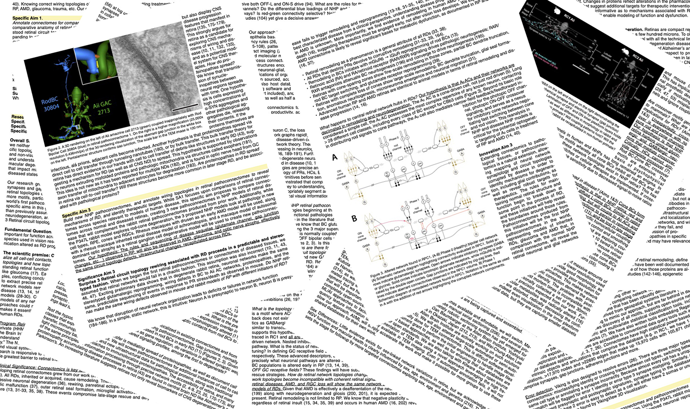
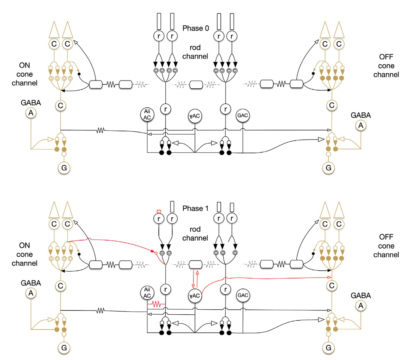
As an undergraduate, I worked with the Biology Department to create some of the world’s first online teaching tools for anatomy and embryology using Hypercard. As an undergrad and then graduate student, I ran some of the first websites and blogs on the Internet from servers here at the University of Utah with the encouragement and blessing of the University of Utah, and have worked with IT over the years to allow this information to be disseminated around the world. I have managed the world’s first online textbook, Webvision for over 25 years that has been hosted on servers in my office or lab, and paid for out of my own pocket. We have modeled Moran CORE on Webvision that is garnering millions of visits per year. We have partnered with the Scientific Computing Institute to create the tools that allowed us to construct the world’s first retinal connectomes. My lab has also created the world’s first pathoconnectomes in any system with software that we have continued to develop, and for many years, we were the only NIH funded laboratory that performed connectomics related work. We have absolutely been global pioneers in this space. This was all possible because the computational environment at the University of Utah allowed and encouraged experimentation and exploration with novel new computational tools.

I will forever be grateful to the people here who helped me along from my chairman, Randy Olson on down.  I will absolutely miss folks here, and it will be bittersweet leaving, but the University of Pittsburgh Ophthalmology program is such an amazing opportunity filled with phenomenal people and goals that I could not resist the offer.  Every year has brought at least one offer to move the lab to another institution, and it’s been easy to turn down many of those offers, but this move to Pitt was the most compelling yet, filled with amazing colleagues who also happen to be good human beings who are engaged in exciting science.  Leadership at Pitt understands that to continue pushing science forward means a commitment to a computational IT environment that is supportive of that work and the role of computational science in it.  They are supportive and eager to grow initiatives there that take advantage of those aspects of biomedicine.  Housing is far more affordable around Pittsburgh than it is here in Salt Lake City, and the vibe in Pittsburgh is one of excitement, equity, and investment in the diverse people that make up the communities around Pittsburgh.  This level of support and commitment to people makes moving the whole lab and all the personnel there much more attractive.

And my vote will count in Pennsylvania.

We will be anchoring histology, ultrastructure and connectomics at Pitt, as well as helping them to hire out the next phase of expansion with sensory neurosciences as a focus.  I’m grateful to José-Alain Sahel, and John Ash for their commitment and efforts to bring us on, and to the entire faculty of the Ophthalmology department, the neurosciences and engineering communities at Pitt who will be our colleagues and collaborators on a variety of science projects moving forward to help understand vision and vision rescues.  As part of this, Becca will be leaving the BWJoneslab team, and taking a completely independent faculty line job with her own startup package at Pitt, so we’ll still be working collaboratively at the same institution which is verrrrry cool.

On a personal note, 2025 has been chaotic and tumultuous for so many reasons. It has been the most difficult, stressful year of my life with changes desired and very much undesired.  This move is a decision that Hilari and I were very much looking forward to as a new adventure for Team Jones, and I am absolutely heartbroken to be making this move without the love of my life and my best friend.  She was looking forward to living in a new city, and all of the excitement of starting fresh and discovering new things, new arts, new opportunities for philanthropy and exploration.  I don’t know how to even process this as it feels like I’ve lost the part of me that helps understand, plan and contextualize everything.  Every move like this has its downsides, but those aspects are the ones that scare me most, and feel the most destabilizing.  I miss you, my love.  And I will continue to work to make you proud of me.

NOA EY028927

I am pleased… no… absolutely relieved to report that we have received the Notice of Award for our NIH Retinal Circuitry grant renewal for EY028927.  Given all that has been going on <gestures widely> with NIH and the federal government, this has been a concern.  We got a very good score on this, Council met back in October, but until now, we’ve not known whether or not this would be awarded, especially with a looming government shutdown.  That makes over a year since the original submission on this.

If it were not for an incredibly generous private donation from Gabe Newell for which I will always be grateful, this past few months would have been tricky indeed.

My thanks to all the peeps at NIH, the grants management specialist, the PO and SRO and all those who labored through uncertain times to push this award through.  You have my undying appreciation.

Excitatory and Inhibitory Neurotransmitter Alterations With Advancing Age and Injury in the Mouse Retina

We have a new paper out in Neurobiology of Aging, Excitatory and Inhibitory Neurotransmitter Alterations With Advancing Age and Injury in the Mouse Retina.

Authors: Katharina C Bell, Vicki Chrysostomou, Markus Karlsson, Bryan William Jones@bwjones.bsky.social, Pete A Williams @petetheteapot.bsky.social, and Jonathan G Crowston

Abstract: Increasing age and elevated intraocular pressure (IOP) are the two major risk factors for glaucoma, the most common cause of irreversible blindness worldwide. Accumulating evidence is pointing to metabolic failure predisposing to neuronal loss with advancing age and IOP injury. Many neurotransmitters are synthesized from endogenous metabolites and are essential for correct cell to cell signaling along the visual pathways. We performed detailed, small molecule metabolomic profiling of the aging mouse retina and further explored the impact of IOP elevation at different ages. The resultant metabolomic profiles showed clear discrimination between young and middle-aged retinas and these changes are accentuated following eye pressure elevation. Alterations in glutamate and Gamma-aminobutyric acid (GABA) related metabolites were the most apparent changes with advancing age with further reductions in GABA and related pathways after IOP elevation. These changes were further confirmed using immunohistochemistry and patch-clamp electrophysiological recording experiments.

Uncovering The Electrical Synapse Proteome In Retinal Neurons Via In Vivo Proximity Labeling

We have a new paper out In BioRxiv, Uncovering The Electrical Synapse Proteome In Retinal Neurons Via In Vivo Proximity Labeling.

Authors: Stephan Tetenborg, Eyad Shihabeddin, Elizebeth Olive Akansha Manoj Kumar, Crystal Sigulinsky @csigulinsky.bsky.social, Karin Dedek, Ya-Ping Lin, Fabio Echeverry, Hannah Hoff, Alberto Pereda, Bryan William Jones @bwjones.bsky.social, Christophe Ribelayga, Klaus Ebnet, Ken Matsuura, John O’Brien

Abstract: Through decades of research, we have gained a comprehensive understanding of the protein complexes underlying function and regulation of chemical synapses in the nervous system. Despite the identification of key molecules such as ZO-1 or CaMKII, we currently lack a similar level of insight into the electrical synapse proteome. With the advancement of BioID as a tool for in vivo proteomics, it has become possible to identify complex interactomes of a given protein of interest by combining enzymatic biotinylation with subsequent streptavidin affinity capture. In the present study, we applied different BioID strategies to screen the interactomes of Connexin 36 (mouse) the major neuronal connexin and its zebrafish orthologue Cx35b in retinal neurons. For in vivo proximity labeling in mice, we took advantage of the Cx36-EGFP strain and expressed a GFP-nanobody-TurboID fusion construct selectively in AII amacrine cells. For in vivo BioID in
zebrafish, we generated a transgenic line expressing a Cx35b-TurboID fusion under control of the Cx35b promoter. Both two strategies allowed us to capture a plethora of molecules that were associated with electrical synapses and showed a high degree of evolutionary conservation in the proteomes of both species. Besides known interactors of Cx36 such as ZO-1 and ZO-2 we have identified more than 50 new proteins, such as scaffold proteins, adhesion molecules and regulators of the cytoskeleton. We further determined the subcellular localization of these proteins in AII amacrine and tested potential binding interactions with Cx36. Of note, we identified signal induced proliferation associated 1 like 3 (SIPA1L3), a protein that has been implicated in cell junction formation and cell polarity as a new scaffold of electrical synapses. Interestingly, SIPA1L3 was able to interact with ZO-1, ZO-2 and Cx36, suggesting a pivotal role in electrical synapse function. In summary, our study provides a first detailed view of the electrical synapse proteome in retinal neurons.

Injury and Repair: Retinal Remodeling for the Encyclopedia of the Eye, 2nd Edition

Screenshot

We’ve got a new chapter out from Bryan Jones and Robert Marc, Injury and Repair: Retinal Remodeling for the 2nd Edition of Encyclopedia of the Eye. (PDF here)

Abstract: Retinal remodeling is a collection of molecular and cellular revisions triggered by retinitis pigmentosa (RP), Usher syndrome, age-related macular degeneration (AMD), and likely glaucoma. These revisions include neuronal rewiring and neurotransmitter receptor reprogramming; neuritogenesis and synaptogenesis (chemical and electrical); self-signaling; neuronal migration; neuronal death; glial hypertrophy; altered glial gene expression; vascular remodeling; retinal pigmented epithelium (RPE) invasion and hyperpigmentation, and in the terminal stages, extensive proteinopathies. Metabolic and circuit revisions begin as soon as photoreceptor stress is initiated, and accelerate upon complete local photoreceptor loss. Retinal remodeling impacts the timing and potential outcomes of gene therapy, survival factor treatments, stem or progenitor cell implantation, retinal transplantation, and bionic implants.

Retinal Connectomics: A Review

We have a new manuscript out of the lab in Volume 10 of the Annual Review of Vision Science titled Retinal Connectomics: A Review by Crystal L. Sigulinsky, Rebecca L. Pfeiffer, and Bryan William Jones. A .pdf is here.

Abstract
The retina is an ideal model for understanding the fundamental rules for
how neural networks are constructed. The compact neural networks of
the retina perform all of the initial processing of visual information be-
fore transmission to higher visual centers in the brain. The field of retinal
connectomics uses high-resolution electron microscopy datasets to map
the intricate organization of these networks and further our understand-
ing of how these computations are performed by revealing the fundamental
topologies and allowable networks behind retinal computations. In this ar-
ticle, we review some of the notable advances that retinal connectomics
has provided in our understanding of the specific cells and the organi-
zation of their connectivities within the retina, as well as how these are
shaped in development and break down in disease. Using these anatomi-
cal maps to inform modeling has been, and will continue to be, instrumental
in understanding how the retina processes visual signals.