Tag Archives: Crystal Sigulinsky

Uncovering the electrical synapse proteome in retinal neurons via in vivo proximity labeling

We have a new paper out in eLife: Uncovering the electrical synapse proteome in retinal neurons via in vivo proximity labeling.

Authors: Stephan Tetenborg, Eyad Shihabeddin, Elizebeth Olive Akansha Manoj Kumar, Crystal Sigulinsky @csigulinsky.bsky.social, Karin Dedek, Ya-Ping Lin, Fabio Echeverry, Hannah Hoff, Alberto Pereda, Bryan William Jones @bwjones.bsky.social, Christophe Ribelayga, Klaus Ebnet, Ken Matsuura, John O’Brien

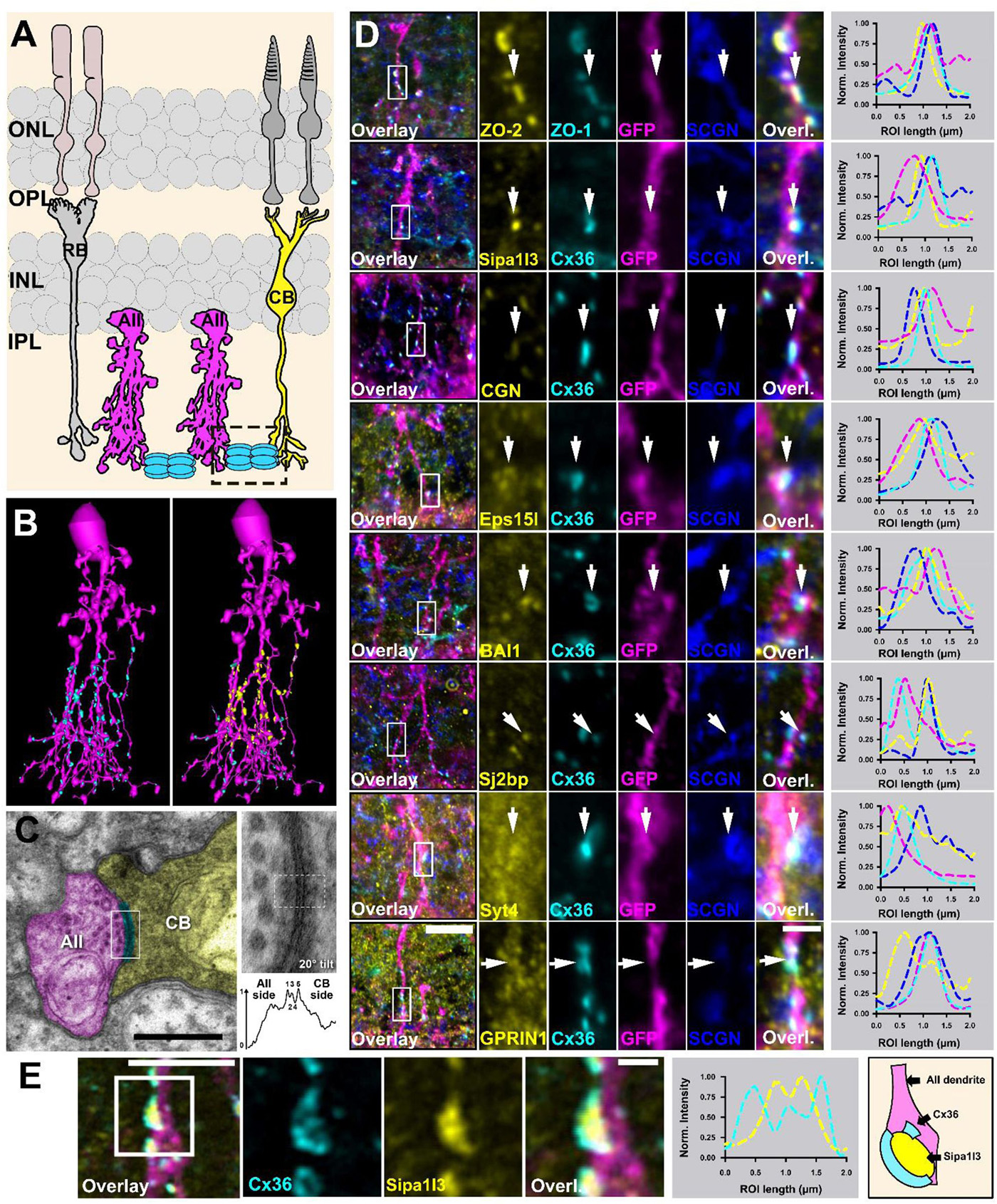
Abstract: Electrical synapses containing Connexin 36 (Cx36) represent the main means for direct electrical communication among neurons in the mammalian nervous system. However, little is known about the protein complexes that constitute these synapses. In the present study, we applied different BioID strategies to screen the interactomes of Connexin 36 and its zebrafish orthologue Cx35b in retinal neurons. For in vivo proximity labeling in mice, we took advantage of the Cx36-EGFP strain and expressed a GFP-nanobody-TurboID fusion construct selectively in AII amacrine cells. For in vivo BioID in zebrafish, we generated a transgenic line expressing a Cx35b-TurboID fusion under control of the Cx35b promoter. Both strategies allowed us to capture a plethora of molecules that were associated with electrical synapses and showed a high degree of evolutionary conservation in the proteomes of both species. Besides known interactors of Cx36 such as ZO-1 and ZO-2 we have identified more than 50 new proteins, such as scaffold proteins, adhesion molecules and regulators of the cytoskeleton. Moreover, we determined the subcellular localization of these proteins in mouse retina and tested potential binding interactions with Cx36. Amongst these new interactors, we identified signal induced proliferation associated 1 like 3 (Sipa1l3), a protein that has been implicated in cell junction formation and cell polarity, as a new scaffold of electrical synapses. Interestingly, Sipa1l3 was able to interact with ZO-1, ZO-2 and Cx36, suggesting a pivotal role in electrical synapse function. In summary, our study provides the first detailed view of the electrical synapse proteome in retinal neurons, which is likely to apply to electrical synapses elsewhere.

Uncovering The Electrical Synapse Proteome In Retinal Neurons Via In Vivo Proximity Labeling

We have a new paper out In BioRxiv, Uncovering The Electrical Synapse Proteome In Retinal Neurons Via In Vivo Proximity Labeling.

Authors: Stephan Tetenborg, Eyad Shihabeddin, Elizebeth Olive Akansha Manoj Kumar, Crystal Sigulinsky @csigulinsky.bsky.social, Karin Dedek, Ya-Ping Lin, Fabio Echeverry, Hannah Hoff, Alberto Pereda, Bryan William Jones @bwjones.bsky.social, Christophe Ribelayga, Klaus Ebnet, Ken Matsuura, John O’Brien

Abstract: Through decades of research, we have gained a comprehensive understanding of the protein complexes underlying function and regulation of chemical synapses in the nervous system. Despite the identification of key molecules such as ZO-1 or CaMKII, we currently lack a similar level of insight into the electrical synapse proteome. With the advancement of BioID as a tool for in vivo proteomics, it has become possible to identify complex interactomes of a given protein of interest by combining enzymatic biotinylation with subsequent streptavidin affinity capture. In the present study, we applied different BioID strategies to screen the interactomes of Connexin 36 (mouse) the major neuronal connexin and its zebrafish orthologue Cx35b in retinal neurons. For in vivo proximity labeling in mice, we took advantage of the Cx36-EGFP strain and expressed a GFP-nanobody-TurboID fusion construct selectively in AII amacrine cells. For in vivo BioID in
zebrafish, we generated a transgenic line expressing a Cx35b-TurboID fusion under control of the Cx35b promoter. Both two strategies allowed us to capture a plethora of molecules that were associated with electrical synapses and showed a high degree of evolutionary conservation in the proteomes of both species. Besides known interactors of Cx36 such as ZO-1 and ZO-2 we have identified more than 50 new proteins, such as scaffold proteins, adhesion molecules and regulators of the cytoskeleton. We further determined the subcellular localization of these proteins in AII amacrine and tested potential binding interactions with Cx36. Of note, we identified signal induced proliferation associated 1 like 3 (SIPA1L3), a protein that has been implicated in cell junction formation and cell polarity as a new scaffold of electrical synapses. Interestingly, SIPA1L3 was able to interact with ZO-1, ZO-2 and Cx36, suggesting a pivotal role in electrical synapse function. In summary, our study provides a first detailed view of the electrical synapse proteome in retinal neurons.

Mitochondrial Transfer Between Inner Retinal Neurons

This abstract was presented today, April 26th at the 2023 Association for Research in Vision and Opthalmology (ARVO) meetings in New Orleans, Louisiana by Selena Wirthlin, Crystal Sigulinsky, James Anderson, and Bryan William Jones.

Full resolution version here.

Purpose
Intercellular mitochondrial transfer has been reported across a variety of cells and tissues under both physiological and pathological conditions. Such transfer has shown broad therapeutic potential. The effectiveness of this therapy, however, is limited by a lack of understanding of the cellular and molecular mechanisms. Here, the ultrastructural features of mitochondrial transfer between inner retinal neurons discovered through retinal connectomics analysis is shown.

Methods
Retinal Connectome 2 (RC2) was built by automated transmission electron microscopy at ultrastructural (2nm/pixel) resolution. RC2 is a 0.25mm diameter volume of retina obtained from a 5-month-old female C57BL/6J mouse. The Viking application was used to visualize and annotate inter- and intracellular features of interest in the connectome.

Results
Exploration of RC2 revealed material transfer between apposing neural processes within the OFF subliminal of the inner plexiform layer. The transferred material can be defined as a mitochondria, confirmed by the presence of crustae. At the transfer site, a short, electron-dense 140-nm diameter tube with a curved cap tightly associated with the inner mitochondrial membrane of one neuritis extends into a vacuole within the apposing neuritis formed by the plasma membranes of the two cells. Thin cytoskeletal components consistent with actin microfilaments extend into the mitochondrion. Morphology and synaptology of the acceptor cell confirm it is an Aii amacrine cell, while preliminary findings suggest the donor cell is a type of ON/OFF ganglion cell.

Conclusions
These findings demonstrate active mitochondrial transfer between different classes of endogenous inner retinal neurons and suggests it may represent an important component of tissue homeostasis in the retina. Features of this transfer differ from previously reported mitochondrial transfer between photoreceptors upon transplantation, which may indicate cell type- or context-dependent differences in the cellular or molecular mechanisms. Our findings demonstrate active mitochondrial transfer between different classes of endogenous inner retinal neurons and suggest it may represent an important component of tissue homeostasis in the retina. Features of this transfer differ from previous reports by the Wallace and Pearson groups of material transfer between photoreceptors upon transplantation through tunneling nanotubes (Ortin- Martinez et al., 2021; Kalargyrou et al., 2021), which may indicate cell type- or context-dependent differences in the cellular or molecular mechanisms. Understanding these mechanisms could serve as a catalyst for development of novel therapeutics for disease in the retina and beyond.

Species-Specific Connectivity In The Aii Connectome

This talk was presented today, April 25th at the 2023 Association for Research in Vision and Opthalmology (ARVO) meetings in New Orleans, Louisiana by Crystal Sigulinsky as part of an ARVO Minisymposium organized by Bryan William Jones.

Abstract: Biomedical research relies heavily on animal models to study human disease and develop therapeutics. Understanding the architectural diversity in neural networks between humans and these model species is essential for choosing a relevant study model and interpreting conflicting results. Using comparative connectomics, we sought to map and compare the local neural network architecture of rabbit and mouse retinal Aii amacrine cells. This specialized narrow-field, multistratified, glycinergic interneuron has critical feedforward and feedback roles in both the photopic and scotopic retinal networks spanning the ON and OFF pathways, making it an ideal candidate for investigating species-specific differences in retinal networks. High-resolution, serial-section transmission electron microscopy (TEM) volumes of rabbit (RC1: female, 13- month, Dutch Belted) and mouse (RC2: female, 5-month, C57BL/6J) retinal tissue provided spatially-registered synaptic maps of Aii connectivity at directly comparable resolution and completeness. These reveal that despite species-specific morphologies, gross synaptology and compartmentalization appear conserved. Yet, rabbit and mouse Aii cells diverge in the weighting of their partnerships, most notably in their coupling profiles. Opposing biases in gap junction partnerships and their respective sizing rules indicate a greater relative output by mouse Aii cells to ON pathways than in rabbit. However, a unique topological conformation for a subset of conventional presynapses formed by Aii cell lobular dendrites with species-specific features and prevalence may influence signal output to specific partner classes within the OFF pathway and either nullify or exacerbate this difference in ON/OFF output. Additionally, rabbit Aii cells in RC1 showed greater Aii-Aii coupling than in mouse, which may suggest greater signal-to-noise compensation. Lastly, preliminary data suggest mouse Aii cells receive greater excitatory, but not inhibitory input/feedback from the OFF pathway than in rabbit. Together these data indicate that precise neural circuit architectures diverge between species and require detailed, comprehensive mapping to begin to dissect potential influence on signal flow.

Structural Motifs Of Excitatory Synapses In The Mammalian Retina

This abstract was presented today, April 24th at the 2023 Association for Research in Vision and Opthalmology (ARVO) meetings in New Orleans, Louisiana by Taylor Otterness, Crystal Sigulinsky, James Anderson, and Bryan William Jones.

Full resolution version here.

Purpose
Connectivity within the nervous system is precise and disruptions lead to degraded performance and disease, yet the rules that govern connectivity remain unknown. Recent efforts reveal that different types of cone bipolar cells in the neural retina show preferences in the selection and frequency of presynaptic structure types used for signal transmission. However, it is not yet known how these differences are related to the quantity or type of postsynaptic partner. We used Retinal Connectome 1 (RC1) to analyze the synaptic output of rabbit CBb6 cells, a type of ON cone bipolar cell that forms excitatory synapses via diverse presynaptic structure types, to identify patterns in how these cells interact with their postsynaptic partners.

Methods
RC1 is a 0.25 mm diameter volume sampled from mid-peripheral retina of a 13 month old female Dutch-Belted rabbit, serially sectioned at 70 nm, and imaged at ultrastructural resolution (2nm/px) using transmission electron microscopy. Postsynaptic partners of CBb6 cell 6156’s presynaptic structures were annotated using the Viking Viewer for Connectomics. Statistical analyses were conducted in Microsoft Excel and investigated further with 3D rendering and graph visualization of connectivity.

Results
The factors tracked for comparison included presynaptic structure type, target number, and postsynaptic partner type. Multiribbon synapses of CBb6 cell 6156 trended towards having a greater number of output partners, with a greater proportion of dyads than monads. Despite this, triads and quadrads were only found opposing single ribbon synapses. As the different presynaptic structure types may differ in the strength of neurotransmitter release (ribbonless < single ribbon < multiribbon), these findings are inconsistent with scaling of output to the number of postsynaptic targets. Both amacrine cells (AC) and ganglion cells (GC) are postsynaptic partners of 6156. However, single ribbon and ribbonless structures appear biased towards AC only targets, while multiribbon synapses appear biased toward mixed AC and GC targets.

Conclusions
Target type relationships appear more important than the number of targets in determining presynaptic structure type in CBb6. Future efforts will examine size differences of postsynaptic structures and presynaptic ribbon size, and even compare across bipolar cell classes, in order to provide further insight on the connectivity rules underlying excitatory synapses.

Marclab Off To ARVO2019

The Marclab is off to ARVO 2019 and eager to share some of what we’ve been up to over the past year.  We have undergraduate Jeebika Dahal presenting her work on the AII Amacrine Cell Connectivity Changes In Degenerating Retina on Sunday (see poster B0013 Abstract Number: 551 – B0013).  Undergraduate Selena Wirthlin will present her work on the Comparative Anatomy and Connectivity Of The AII Amacrine Cell In Mouse And Rabbit Retina on Sunday (poster B0010 Abstract Number: 548 – B0010). Undergraduate and US Navy veteran Jessica Garcia will present her work Sunday on OFF-layer Branches Of ON Cone Bipolar Cells In Early Retinal Degeneration (B0017 Abstract Number: 555 – B0017). And postdoc Crystal Sigulinsky will present her work on Coupling Architecture Of The Aii/ON Cone Bipolar Cell Network In Degenerate Retina in a platform presentation on Thursday at 11:15am (Abstract Number: 6441).

We hope to see you there!

Off to RD2018 and ISER 2018

The Marclab for Connectomics is off to RD2018 and ISER 2018 in Killarney, Ireland and Belfast, Northern Ireland.  I’ll be organizing sessions on retinal degeneration, and I’m tremendously proud of the work Dr. Crystal Sigulinsky will be presenting from her work on gap junctional connectivity in retinal degenerations and the work Dr. Rebecca Pfeiffer (@BeccaPfeiffer19) will be presenting on her work on the retinal pathoconnectome in two talks on bipolar cells and Müller cells.

Synaptic Inputs To A Gamma Ganglion Cell In Rabbit Retina

This abstract was presented today, May 8th at the 2017 Association for Research in Vision and Opthalmology (ARVO) meetings in Baltimore, Maryland by Andrea Bordt, Diego Perez, Robert E. Marc, James R. Anderson, Carl B. Watt, Bryan W. Jones, Crystal Sigulinsky, James S. Lauritzen, Danny Emrich, Noah Nelson, Luke Tseng, Weiley Liu, and David W. Marshak. Full resolution version here.

Purpose: There are at least 30 distinct types of mammalian retinal ganglion cells, each sensitive to different features of the visual environment, and these can be grouped according to their morphology. One such group, the gamma cells, was identified more than 40 years ago, but their synaptic inputs have never been described. That was the goal of this study.

Methods: The synaptic inputs to a subtype of gamma cell with dendrites ramifying in the outer sublamina of the inner plexiform layer (IPL) of the rabbit retina were identified in a retinal connectome developed using automated transmission electron microscopy.

Results: The gamma cell was always postsynaptic in the IPL, confirming its identity as a ganglion cell. The local synaptic input should produce relatively weak OFF reposnses to stimuli confined to the center of the gamma cell’s receptive field. It typically received only one synapse per bipolar cell from at least 4 types of OFF bipolar cells. Because bipolar cells vary in their response kinetics and contrast sensitivity. each type would provide a small, asynchronous excitatory input. The amacrine cells at the dyad synapses provided only a small amount presynaptic inhibition; reciprocal synapses were observed in only 3 of the 18 ribbon synapses. There was no glycinergic crossover inhibition, another local interaction that would enhance light responses. Local postsynaptic inhibition was somewhat more common; in 6 instances, the bipolar cells presynaptic to the gamma cell or their electrically coupled neighbors also provided input to an amacrine cell that inhibited the gamma cell. The other amacrine cell inputs to the gamma cell should have a much greater impact on the light responses because they are far more numerous. These are from axons and long dendrites of GABAergic amacrine cells, and they provide 60% of all the input. This finding suggests that many types of stimuli in the receptive field surround or outside of the classical receptive field would provide potent inhibition to the gamma cell.

Conclusions: The synaptic inputs rsuggest that gamma cells in rabbit retina would have light responses like their homologs in mouse retina, OFF responses to small stimuli in the receptive field center that are suppressed by a variety of larger stimuli. Thus, they would signal object motion selectively.