We finished sectioning and capturing a massive new retinal connectome that we are going to be so excited to announce at some point in the not too distant future. Effective immediately, we are also starting on a brand new pathoconnectome that we will be powering through over the next little while. Thanks to the team of people who make this happen, shown in this post are Jia-Hui Yang, Matt Berardy, and Rebecca Pfeiffer. More photos here.
Category Archives: Connectomics
FASEB Retina Meeting 2022
Lab PI Bryan Jones and postdoctoral fellow Crystal Sigulinsky presented work at the FASEB Retina Meeting, 2022 in Southbridge, Massachusetts. Edit: Photos of the meeting can be seen here.
Seminar: Retinas, How Are They Wired? How Does Wiring Change In Disease?
PI, Bryan William Jones delivered an invited seminar today at the National Institutes of Health (NIH) / National Eye Institute (NEI) today on Retinas, How Are They Wired? How Does Wiring Change In Disease?
Seminar: Tools and Approaches for Assembly, Review, and Analysis of Large-Scale Electron Microscopy
PI, Bryan William Jones delivered a seminar at the Microscopy and Microanalysis 2021 meeting today on Tools and Approaches for Assembly, Review, and Analysis of Large-Scale Electron Microscopy.
Model-Based Comparison of Current Flow in Rod Bipolar Cells of Healthy and Early-Stage Degenerated Retina
We have a new manuscript out in Experimental Eye Research, Model-Based Comparison of Current Flow in Rod Bipolar Cells of Healthy and Early-Stage Degenerated Retina. (pdf here)
Authors: Pragya Kosta, Ege Iseri, Kyle Loizos, Javad Paknahad, Rebecca L. Pfeiffer @BeccaPfeiffer19, Crystal L. Sigulinsky @CLSigulinsky, James R. Anderson, Bryan W. Jones @BWJones, and Gianluca Lazzi.
Abstract: Retinal degenerative diseases, such as retinitis pigmentosa, are generally thought to initiate with the loss of photoreceptors, though recent work suggests that plasticity and remodeling occurs prior to photoreceptor cell loss. This degeneration subsequently leads to death of other retinal neurons, creating functional alterations and extensive remodeling of retinal networks. Retinal prosthetic devices stimulate the surviving retinal cells by applying external current using implanted electrodes. Although these devices restore partial vision, the quality of restored vision is limited. Further knowledge about the precise changes in degenerated retina as the disease progresses is essential to understand how current flows in retinas undergoing degenerative disease and to improve the performance of retinal prostheses. We developed computational models that describe current flow from rod photoreceptors to rod bipolar cells
Model-based Comparison of Current Flow in Rod Bipolar Cells of Healthy and Early-Stage Degenerated Retina 
Chapter: Retinal Connectomics
We have a new chapter out in the Elsevier book series The Senses, 2021.
Authors are myself, Bryan W. Jones @BWJones.bsky.social and Robert E. Marc.
Abstract: The retina is both a light sensor and a highly complex image-processing device – like supercomputers at the backs of eyes. The retina is also wonderfully compact with all circuitry (glia, neurons, synapses and gap junctions) required to compute sensory input, making it a convenient model for understanding the rest of the nervous system. This is also true for disease, with early evidence indicating retina may be a good model for studying progressive neural degenerative diseases. Modern ultrastructural approaches to the study of neural connections is a relatively new !eld has been termed “connectomics”. Connectomics approaches applied to the retina is termed retinal connectomics. These approaches are relatively new !elds that leverage modern technologies in light and ultrastructural imaging, computational storage, and data management to allow tracking of neuronal identity and connectivity, delivering a robust edge/node network map of circuit topologies. Understanding circuit topologies is critical to understanding how retinas process information, and how information processing is corrupted in disease. This chapter summarizes early history, discusses technical aspects of imaging connectomes, justi!es the importance of why connectomics approaches are important, particularly in retina, discusses what has been learned from early efforts in connectomics, and points the way to the next steps.
Please email me: bryan.jones@pitt.edu if you would like a pdf of the chapter.
A pathoconnectome of early neurodegeneration: Network changes in retinal degeneration
We have a new manuscript out in Experimental Eye Research, A pathoconnectome of early neurodegeneration: Network changes in retinal degeneration. (pdf here)
Authors: Rebecca L. Pfeiffer @BeccaPfeiffer19, James R. Anderson, Jeebika Dahal, Jessica C. Garcia, Jia-Hui Yang, Crystal L. Sigulinsky @CLSigulinsky, Kevin Rapp, Daniel P. Emrich, Carl B. Watt, Hope AB Johnstun, Alexis R. Houser, Robert E. Marc @robertmarc60, and Bryan W. Jones @BWJones.
Abstract: Connectomics has demonstrated that synaptic networks and their topologies are precise and directly correlate with physiology and behavior. The next extension of connectomics is pathoconnectomics: to map neural network synaptology and circuit topologies corrupted by neurological disease in order to identify robust targets for therapeutics. In this report, we characterize a pathoconnectome of early retinal degeneration. This pathoconnectome was generated using serial section transmission electron microscopy to achieve an ultrastructural connectome with 2.18nm/px resolution for accurate identification of all chemical and gap junctional synapses. We observe aberrant connectivity in the rod-network pathway and novel synaptic connections deriving from neurite sprouting. These observations reveal principles of neuron responses to the loss of network components and can be extended to other neurodegenerative diseases.
NeuroNex Grant
I am pleased to report that the The Marclab for Connectomics has been funded by the National Science Foundation with a 5 year grant as part of a large, international consortium to study synaptic weighting. We are collaborating with the Erik Jorgensen laboratory here at the University of Utah, and will be exploring synapses in a model of retinal degeneration. There is a nice writeup of the award on the Moran Eye Center website, here.
This is a wonderful opportunity to work with other colleagues that will be funded alongside us with this grant, including Uri Manor @manorlaboratory, Davi Bock @dddavi, Josh Vogelstein @neuro_data, Viren Jain @stardazed0, and others. My thanks to Kristen Harris for heading up this initiative.
Network Architecture of Gap Junctional Coupling among Parallel Processing Channels in the Mammalian Retina
We have a new manuscript out in The Journal of Neuroscience, Network Architecture of Gap Junctional Coupling among Parallel Processing Channels in the Mammalian Retina.
Authors: Crystal L. Sigulinsky @CLSigulinsky, James R. Anderson, Ethan Kerzner @EthanKerzner, Christopher N. Rapp @ChrisNRapp, Rebecca L. Pfeiffer @BeccaPfeiffer19, Taryn M. Rodman, Daniel P. Emrich, Kevin D. Rapp, Noah T. Nelson @nooneelseinhere, J. Scott Lauritzen, Miriah Meyer@miriah_meyer, Robert E. Marc @robertmarc60, and Bryan W. Jones @BWJones.
Abstract: Gap junctions are ubiquitous throughout the nervous system, mediating critical signal transmission and integration, as well as emergent network properties. In mammalian retina, gap junctions within the Aii amacrine cell-ON cone bipolar cell (CBC) network are essential for night vision, modulation of day vision, and contribute to visual impairment in retinal degenerations, yet neither the extended network topology nor its conservation is well established. Here, we map the network contribution of gap junctions using a high-resolution connectomics dataset of an adult female rabbit retina. Gap junctions are prominent synaptic components of ON CBC classes, constituting 5%–25% of all axonal synaptic contacts. Many of these mediate canonical transfer of rod signals from Aii cells to ON CBCs for night vision, and we find that the uneven distribution of Aii signals to ON CBCs is conserved in rabbit, including one class entirely lacking direct Aii coupling. However, the majority of gap junctions formed by ON CBCs unexpectedly occur between ON CBCs, rather than with Aii cells. Such coupling is extensive, creating an interconnected network with numerous lateral paths both within, and particularly across, these parallel processing streams. Coupling patterns are precise with ON CBCs accepting and rejecting unique combinations of partnerships according to robust rulesets. Coupling specificity extends to both size and spatial topologies, thereby rivaling the synaptic specificity of chemical synapses. These ON CBC coupling motifs dramatically extend the coupled Aii-ON CBC network, with implications for signal flow in both scotopic and photopic retinal networks during visual processing and disease.
Pathoconnectome Analysis of Müller Cells in Early Retinal Remodeling
We have a new manuscript out in Clinical Neurophysiology, An Update on Retinal Prostheses. PubMedDirect Link PDF here.
Authors: Rebecca L Pfeiffer, James R Anderson, Daniel P Emrich, Jeebika Dahal, Crystal L Sigulinsky, Hope AB Morrison, Jia-Hui Yang, Carl B Watt, Kevin D Rapp, Mineo Kondo, Hiroko Terasaki, Jessica C Garcia, Robert E Marc, and Bryan W Jones.
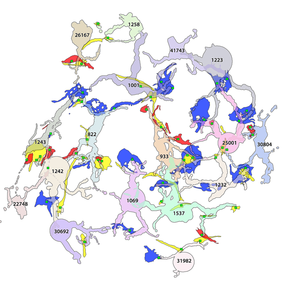
Abstract: Glia play important roles in neural function, including but not limited to amino acid recycling, ion homeostasis, glucose metabolism, and waste removal. During retinal degeneration and subsequent retinal remodeling, Müller cells (MCs) are the first cells to show metabolic and morphological alterations in response to stress. Metabolic alterations in MCs chaotically progress in retina undergoing photoreceptor degeneration; however, what relationship these alterations have with neuronal stress, synapse maintenance, or glia-glia interactions is currently unknown. The work described here reconstructs a MC from a pathoconnectome of early retinalremodeling retinalpathoconnectome 1 (RPC1) and explores relationships between MC structural and metabolic phenotypes in the context of neighboring neurons and glia. Here we find variations in intensity of osmication inter- and intracellularly, variation in small molecule metabolic content of MCs, as well as morphological alterations of glial endfeet. RPC1 provides a framework to analyze these relationships in early retinal remodeling through ultrastructural reconstructions of both neurons and glia. These reconstructions, informed by quantitative metabolite labeling via computational molecular phenotyping (CMP), allow us to evaluate neural-glial interactions in early retinal degeneration with unprecedented resolution and sensitivity.


